Nikon Celebrates 101 Years of Microscopes With Look at the Hidden World of Photomicrography
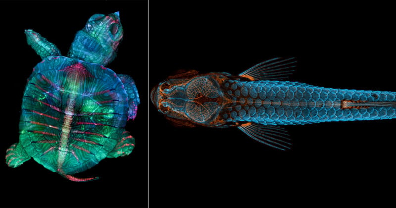
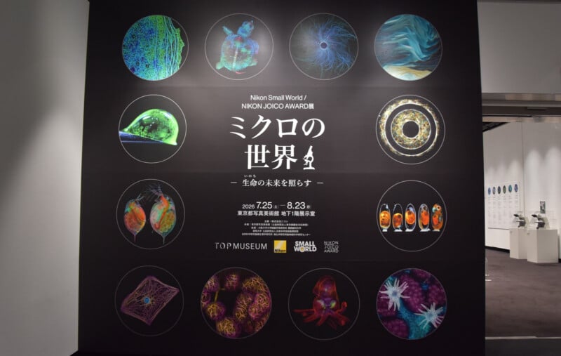
Nikon’s microscope business is celebrating its 101st anniversary this year, and Nikon is marking the occasion with a special exhibition at the Tokyo Photographic Art Museum.
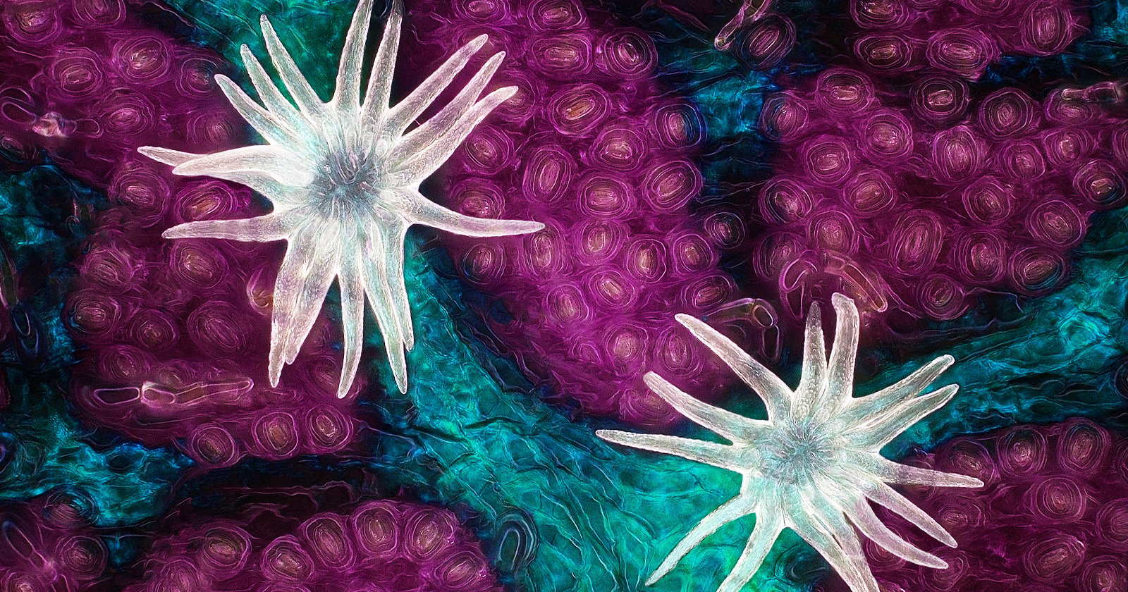
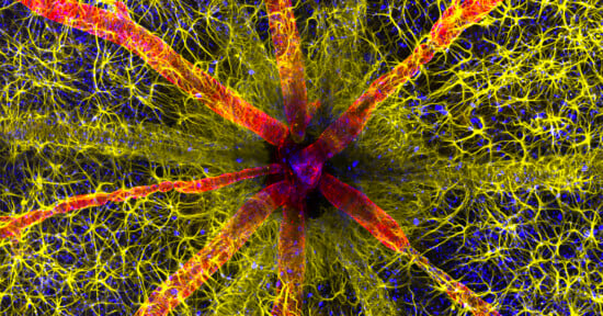
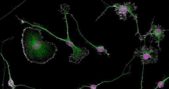
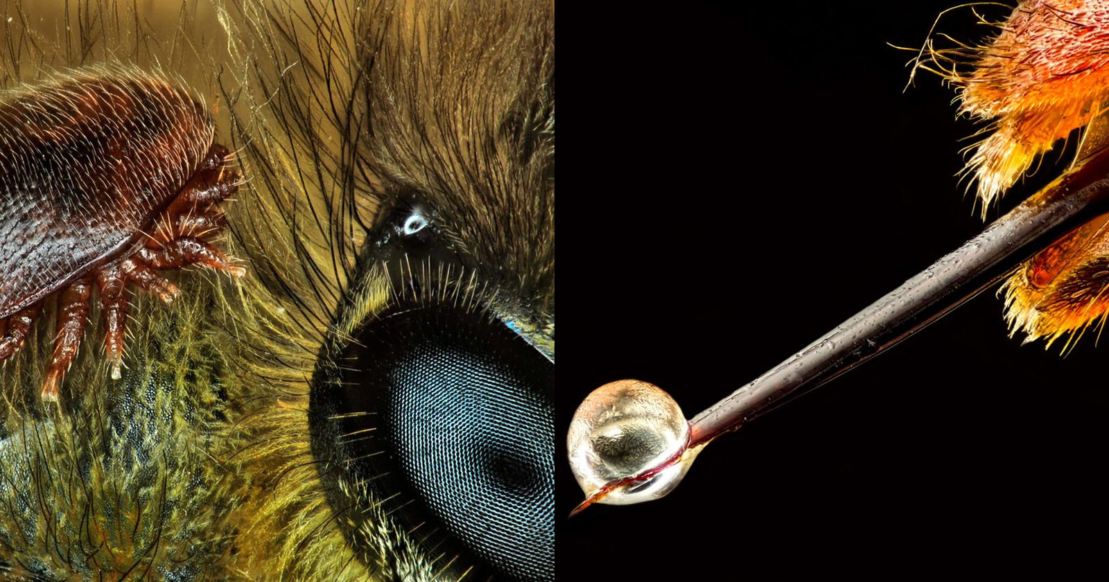
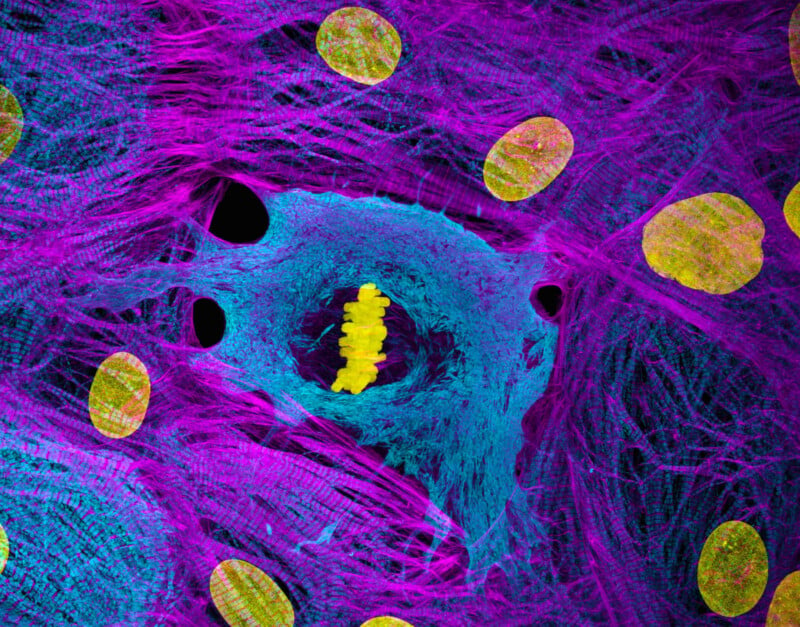
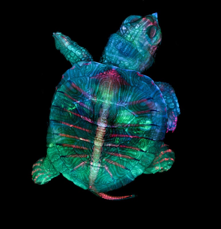
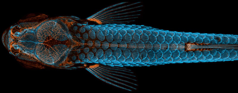
The exhibition, Nikon Small World / Nikon JOICO Award, the Microscopic World — Accelerating Life Science With the Power of Light, primarily features award-winning works from Nikon’s celebrated international photomicrography competitions, like last year’s 51st Nikon Small World competition.
-1-800x533.jpg)
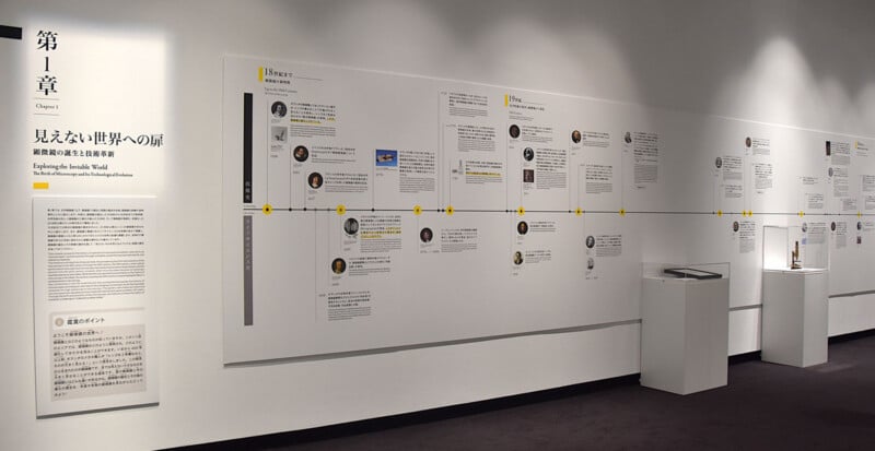
Alongside these beautiful photos, the exhibition also features historical microscopes and works from Nikon’s and the Tokyo Photographic Museum’s collections. There are also areas dedicated to the various use cases of microscopy, including science, medicine, and industry.
Photomicrography is not just beautiful to look at, it is often part of cutting-edge scientific and biomedical research. Many of the award-winning photos in Nikon’s Small World contest were created as part of academic and professional projects. Nikon notes that its new Small World exhibition will celebrate both the artistry and technical expertise of photomicrography.
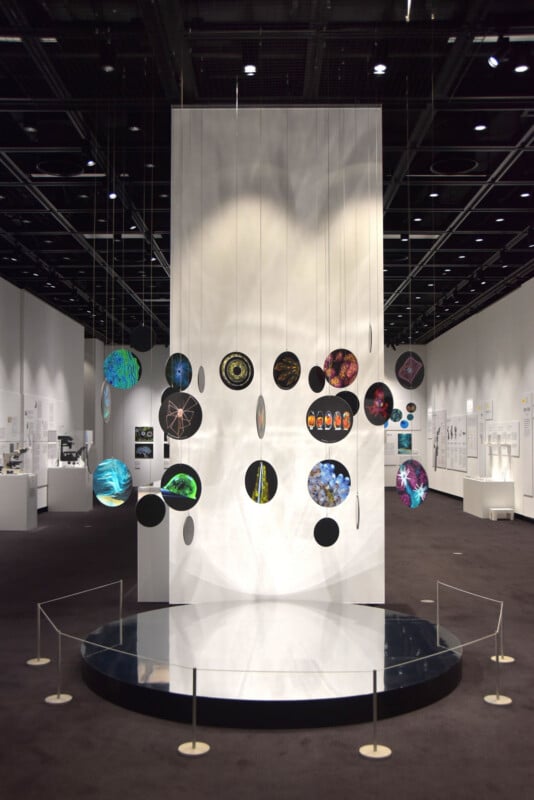
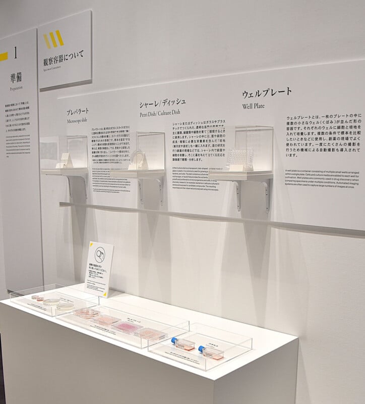
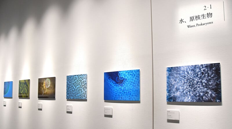
Nikon’s The Microscopic World exhibit includes three primary areas: Gateway to the Invisible World, The Microscopic World, and The Worlds Opened by Microscopes.
Visitors will start with the history of the microscope, from its origins to Nikon’s first microscope from nearly a century ago.
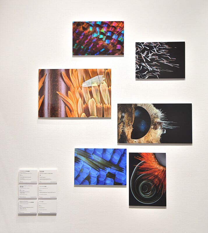
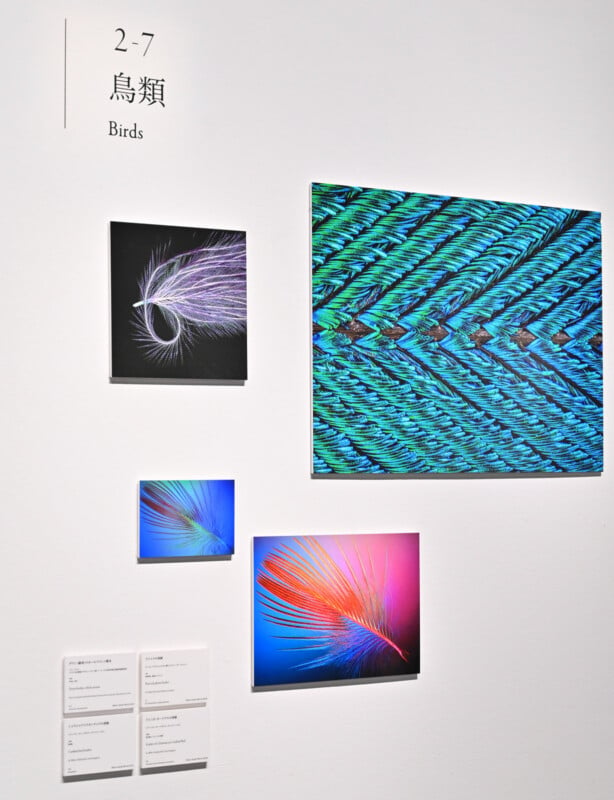
Then they will get to experience recent award-winning works from the Nikon Small World and Nikon JOICO Award competitions, arranged in order of the origins and evolution of life.
“Through photographs and video works, visitors can enjoy the appeal of a microscopic world where science and art come together,” Nikon explains.
Finally, the third section offers educational insights into how microscopes are used to enrich people’s lives and help them, including how people use microscopes in medicine and science. Visitors will also be able to look through a variety of microscopes, which should prove especially exciting to children who have never seen the microscopic world before.
The exhibition started on July 25 and runs through August 23 at the Tokyo Photographic Art Museum in Meguro-ku, Tokyo. Young students and children can visit for free, while adults pay a small fee, around $4. Additional information is available on Nikon’s website and on the Tokyo Photographic Art Museum website.
Image creditsNikon. Individual photographers credited in the captions.